Simple Technique to Improve Cosmetics Post Enucleation
Most of us will do an enucleation or several during our careers, whether you are a general practitioner, ER vet or surgery specific. Here are some tips to improving outcomes and cosmeses.
*Prior to surgery after the induction of anesthesia, a retrobulbar bupivicaine (or lidocaine) block can be administered.
*The key to enucleation include removing all of the conjunctival tissue. The medial canthal region takes extra time and effort to remove all of the firm attachments to the bone. If tissue is missed in this region, a non-healing wound with occur and require additional surgery.
* When dissecting around the globe, avoid pulling the globe, which in turn will pull the optic nerve both ipsilaterally and contralaterally, potentially resulting in blindness in remaining eye.
*I do not typically ligate the optic artery. Instead, I typically apply mild pressure until hemorrhage stops. I find a gelatin sponge (gel-foam or similar, dental squares are a convenient size) place in the region helps provide short and long term hemorrhage control.
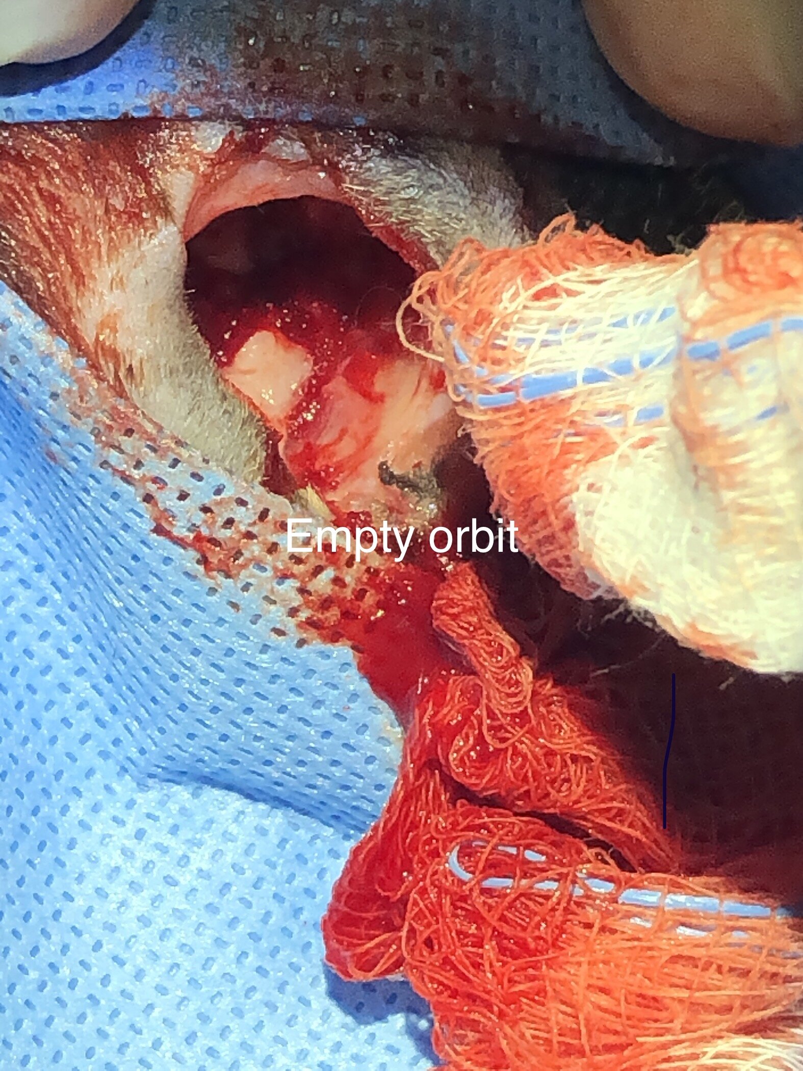
*Prior to tissue closure, I place subperiosteal or at least in the deep tissue of the orbit in a grid-like or zig-zagged fashion. This allows for a platform for the soft tissue closure, avoiding the sunken eye look. A prosthesis can be placed as well, if available and desired.
Subperiosteal sutures are zig-zagged across the empty globe. This provides a scaffold for the subcutaneous tissue to avoid a concave appearance long term. A longer lasting suture is selected such as 3.0 PDS in this illustration.
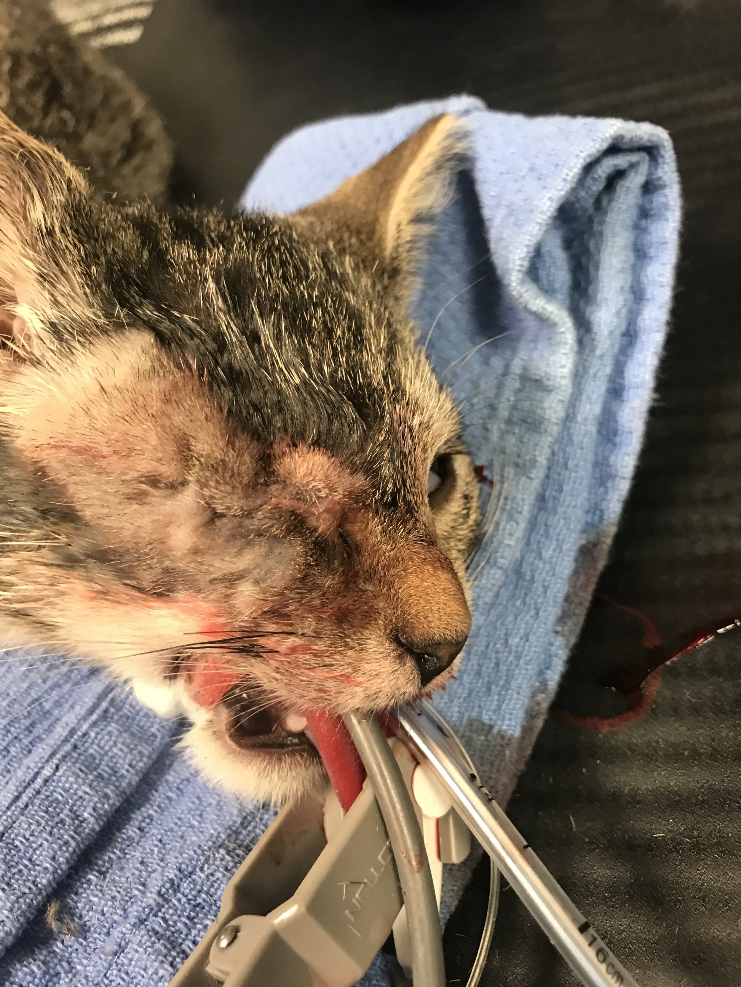
Immediate post operative view of completed enucleation with scaffold in place below.

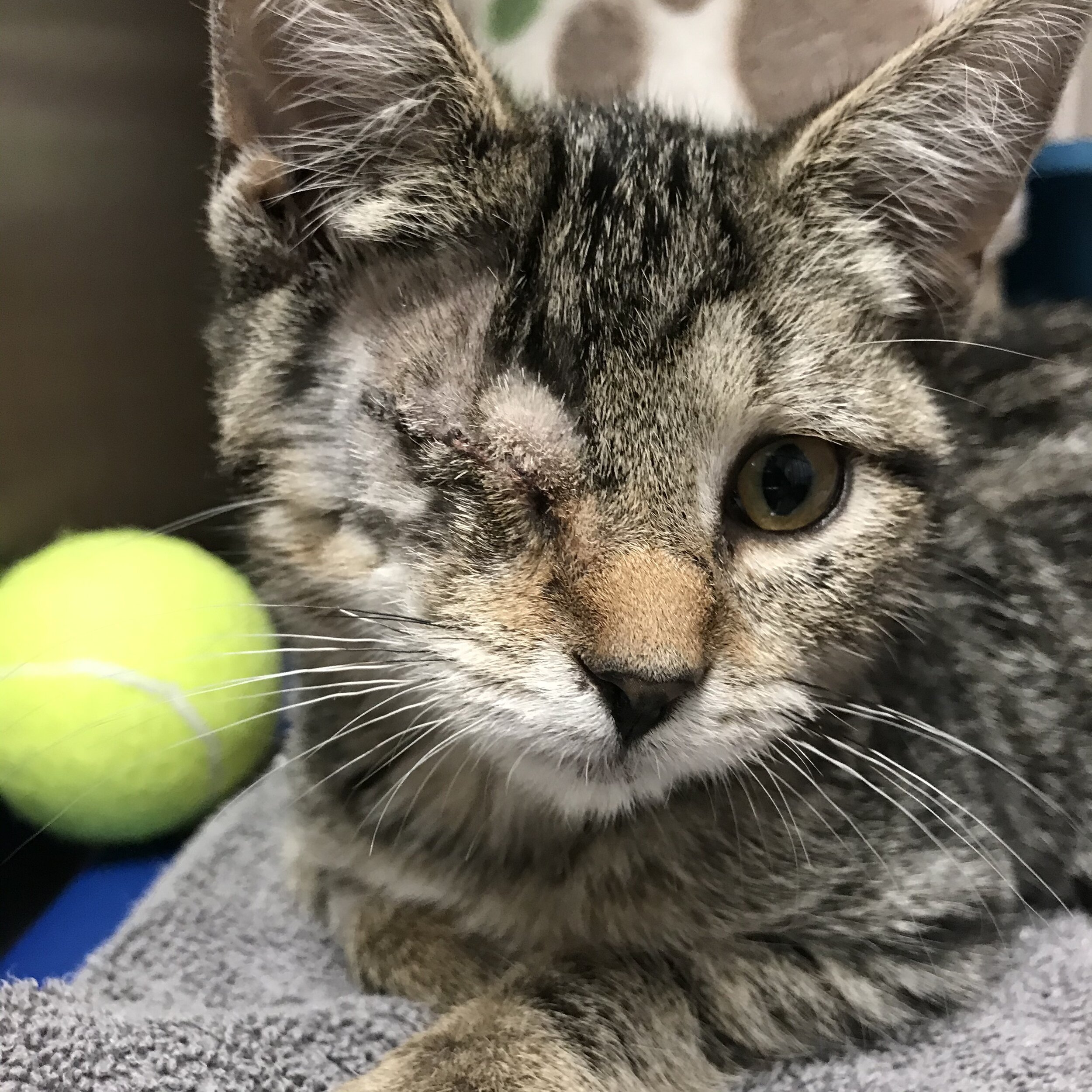
Here is the same cat from previous images, well-healed approximately 3 months later. Note the pleasant closed eye look.
