Medial Patellar Luxation in Cats: Keeping MPLs on your radar for feline patients.

Medial patellar luxation (MPL) is most commonly appreciated in dogs. Cats, however, can have debilitating lameness from medial patellar luxation. Unlike dogs, some cases feline cases can resolve their lameness without surgery. Higher grade luxations may do better with surgical correction. Persistent lameness is an indication for surgery. Cats can do very well with surgical correction with similar techniques used dogs.
Feline patellar luxation is a developmental disease that is more frequently seen in certain breeds such as Manx and Devon Rex. The trochlear ridge forms with persistent motion of the patella in the central trochlear region. If pelvic limb and quadriceps mechanism malaligned for any reason, the patella may not track properly during formation of the groove resulting in patellar luxation. Below is a video of a 15 week old cat with bilateral MPLs. Note the hunched back and "cowboy" style gate. This cat has grade IV/IV MPLs.
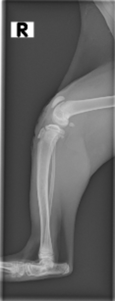
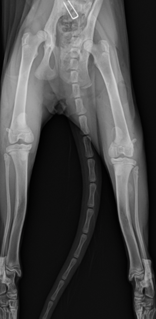
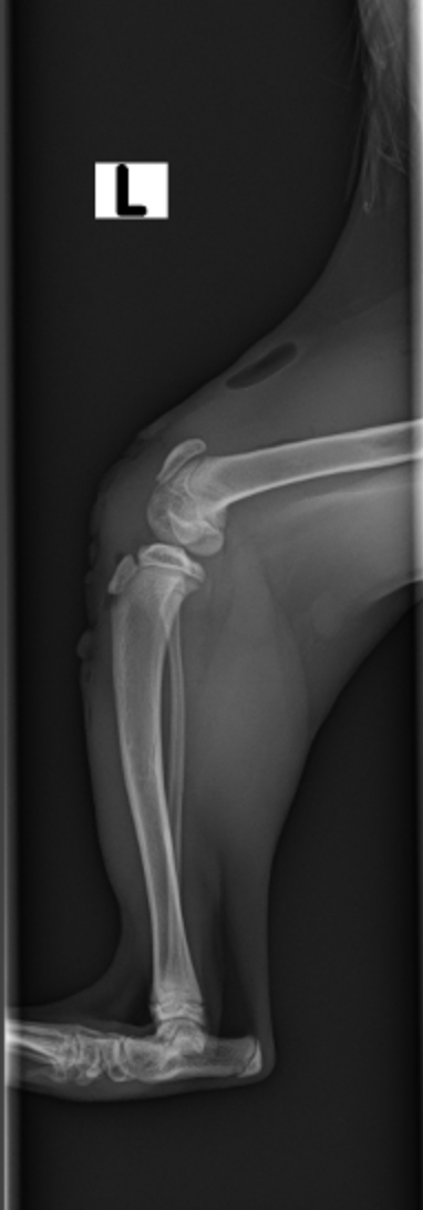
The radiographs (x-rays) from this cat document the grade IV/IV medial patellar luxation condition, both in the VD and lateral views. Note that the patellae are medial to the grooves in the VD. In the lateral view, the patella is noted alongside the trochlear groove, overlapping the groove due to its luxation.


This is a ventrodorsal radiographic view of the pelvics and knees of a cat with grade IV/IV MPLs. The kitten is 15 weeks old. Note medial placement of patellae.

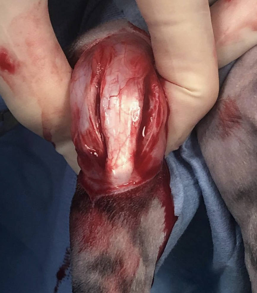
This is an intraoperative picture of a kitten (carniomedial approach) with grade four medial patellar luxations. Note the patella is to the left which is medial. To the far right is the trochlear groove. The entire quadriceps mechanisms is being pulled medially.

First step after medial arthrotomy is to release the retinaculum medially. The release goes all the way through the joint capsule and continues until the joint is visualized. This releases the tightening tissue on the medial aspect. In severe cases, such as this kitten, this release is extended up proximally to release the quadriceps.
After the medial release and arthrotomy, the lateral aspect of the stifle joint is addressed. Redundant lateral retinaculum is excised and the remaining cut edges are imbricated using a pattern such as vest-over-pants. This is to pull the patella laterally into the trochlear groove.

The lateral retinaculum is imbricated and the medial aspect is typically not closed, or closed with the intention of not over tightening the medial aspect.

The end result of soft tissue surgical correction should have the patella in the trochlear groove. The subcutaneous tissues and skin layers are then closed. Generally the medial retinaculum is left open and released.
Immediately post-operatively, radiographs may be taken to verify the placement of the patella(e) in the proper position. Note patellae are in place on the lateral views and more central in the cranial caudal hip views.
At the two week recheck examination, improvement is noted; however posture remains hunched and the legs are still somewhat flexed. Patellae are intermittently in and out of the joint (grade II-III/IV). Strict cage rest is continued. Owners note improvement at home restricted to a large crate.
At the six weeks, marked improvement is noted. Posture is improved. The back is straighter and the pelvic limbs are more naturally extended. Ambulation occurs with more ease. Next recheck scheduled for 10 weeks. Continued cage rest.