Episioplasty: the surgical helper for chronic urinary tract infections in young dogs.
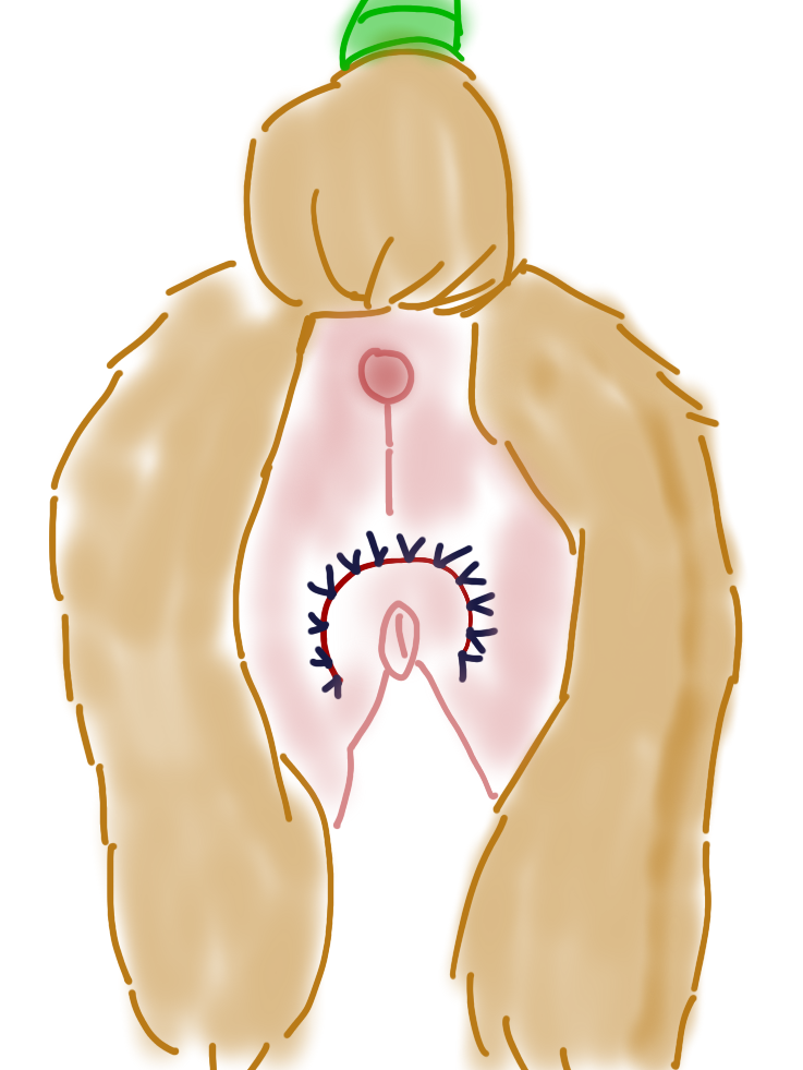
Chronic urinary tract infections in juvenile or younger patients can be the result of a recessed vulva. Obese dogs may also develop vulvar folds. The vulvar folds create a moist environment with recessed regions to trap vulvar and urinary secretions. Ultimately, infection can result both of the skin and the urinary tract. Removing the skin folds surgically in juvenile dogs with recessed vulvas or weight-loss in the obese patient can resolve recurrent infections.
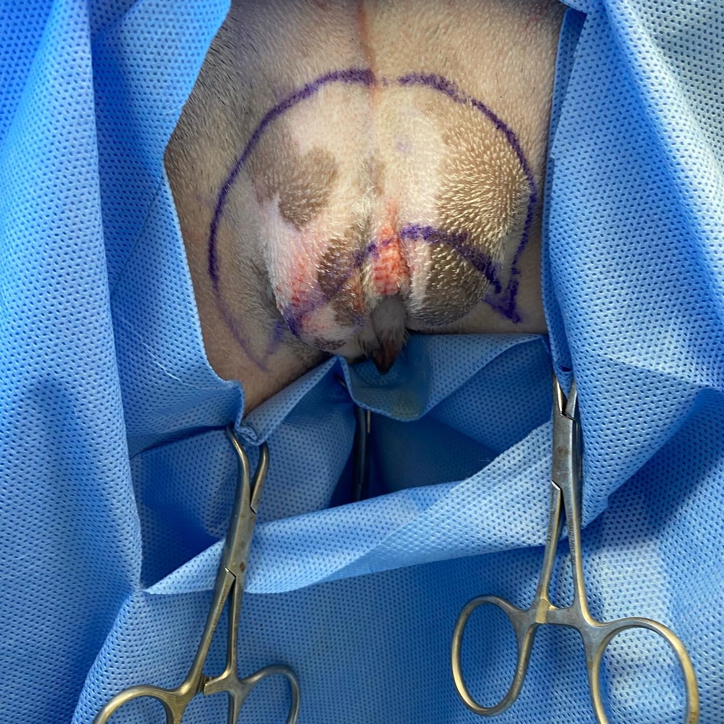
Pictured above is a 2 year old female intact St. Bernard. She presented with a history of urinary tract infections and the need for a spay/gastropexy procedure. She was a fit dog with a body condition of 5/9 yet had a significant vulvar fold. For surgery, she was placed in sternal recumbency. Her legs were placed hanging off the end of the surgery table. Towels were placed on the table end to pad her thighs and protect her femoral nerves from neuropraxia. THIS IS VERY IMPORTANT.**
First, I pinch the tissue above the vulva that I intend to remove. Next, I use a surgical marker to draw the outlines of the tissue to be removed. This is a helpful tip for achieving a symmetrical and effective excision of vulvar fold tissue. Markers can be purchased from most major veterinary distributors.

Sterile Surgical Marker
The surgical markers are sold as presterilized individual units that include a paper ruler. They are a one time use. They can be useful for skin and reconstruction surgeries of all types.
https://www.pdchealthcare.com/sterile-skin-marker-pen-with-flexible-ruler-100-cs-ster-skm.html
Next, the tissue is excised by a combination of blade and scissor excision. Although, a 10 bade is less traumatic, scissors (Mayos) can be more maneuverable in some cases. The skin is then excised along with some subcutaneous tissue. Management of hemostasis is helpful for visibility during surgery and to help minimize hematoma formation and excessive bruising post operatively. Take care not to dissect deeply as the urethra lies below the subcutaneous fat. If in doubt, place a urinary catheter as an anatomic aid. There is a midline fascia layer that is tough and can be worrisome of the urethra but in fact is not. Placing urinary catheters the first few times can help the surgeon to get an idea of the depth of the urethra.

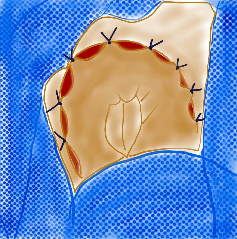
Note the large “moon-shaped” incision made in the perivulvar skin. Adequate skin must be removed to allow for resolution of clinical signs.

Once the tissue is excised, initial tacking sutures are placed at the midline region and bisecting the remaining space. Once the placement is acceptable and symmetrical additional interrupted deep subcutaneous sutures may be placed. I typically use 3.0 PDS or Monocryl.

The sutures placed in each of the following pictures is placed as a “skin suture” for visibility and illustrative purposes. In reality, these sutures are placed subcutaneously.
A continuous layer is then placed in the superficial subcutaneous region and intradermal regions. Based on the size of the dog, I use 3.0 or 4.0 Monocryl (poligecaprone ) or similar. It is my preference to place skin sutures (3.0 ethilon) as an added layer of security as well as improving cosmesis with precise skin apposition.

Dog ears can easily form at the ventral aspects of the incisions. These are easily remedied by the routine tricks. In this particular case, a dog ear was removed from the right side of the incision by cutting out a triangle of tissue.

I leave the sutures tags long for an easier removal. In this case 3.0 ethilon (nylon, ethicon.com) was used in interrupted fashion.

Here is the immediate post operative view of the episioplasty on the St. Bernard.
Post operatively, owners should be warned that bruising is marked to severe. Bruising can occur along the incision as well as in the local skin of the thigh region. As long as no drainage is noted even severe bruising can be normal. In this case we used Nocita (extended-release liposomal bupivacaine). Nocita has a 3 day duration which is ideal for this region with such extensive nerve supply and thus ability to be painful.

This is the two week post operative recheck of the St. Bernard. Note the resolution of bruising, and the continued visibility of the vulva.
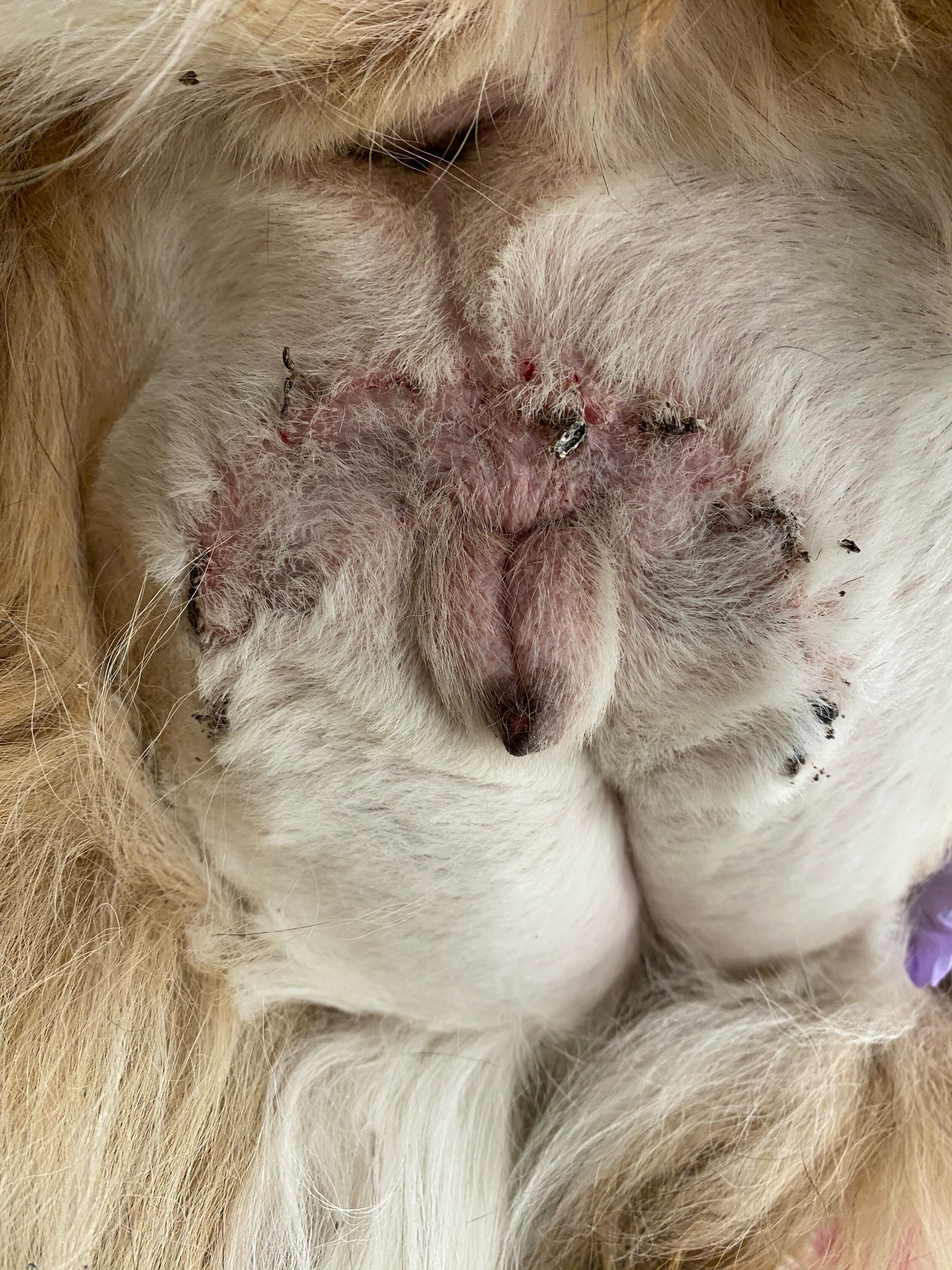
After suture removal, mild scabbing may be noted. At this point a urine sample should be rechecked to ensure bacterial infection has resolved if previously present.